Noone is MRI radiologist par excellence
by Cindy AbolePublic Relations
She's a renaissance woman.
As an MUSC radiologist, Tara Noones focus may seem all science and technology. But it's her love for the English language and the written word and her other creative interests that offer a distinctive balance of skill and flair to this Charleston-born physician.
 Dr.
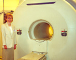
Noone beside one of two of radiology's MRI units.
Dr.
Noone beside one of two of radiology's MRI units.
Noone is the newest specialist to join the Department of Radiology as Assistant Professor of Radiology and Director of Magnetic Resonance Imaging (MRI).
As part of medicines technically-exploding fields, Noone works with a skilled team of radiology technologists, radiologists and a resident-in-training to deliver the highest quality of clinical MRI services.
But Noones start in medicine and radiology was not so direct. She never seriously considered medicine as her ultimate field of study but believed others thought more so.
I knew I liked the sciences but I was also attracted to writing and the telecommunications, radio and television, recalls Noone, whose early experiences as a Notre Dame undergrad attracted her to broadcast journalism working as a radio news reporter, program director and, ultimately, station manager. She also dabbled in television working as a television health news anchor and editor at New York Citys SIC-TV. Medicine was just a logical way to combine all of my interests. I had always wanted to do some type of service work. Through medicine I felt that I could do some good, as well as pursue my own interests.
Noones primary goal is to bring MUSC's MRI Imaging Center up to a state-of-the-art imaging statussimilar in caliber to other top notch MRI centers and radiology programs across the country.
We were fortunate at Chapel Hill to have the capability of providing the most updated MRI services for patients, said Noone, who completed an abdominal MRI fellowship at the University of North Carolina (UNC) hospitals in Chapel Hill. The equipment and technology had been well-established, so clinicians felt comfortable utilizing its capabilities. Here at MUSC, its capabilities are not used enough.
Noone would like to change that. She has also been appointed by radiology to serve as a liaison and imaging specialist with MUSCs Digestive Disease Center (DDC). In addition to her radiology work, Noone will also share her imaging expertise, teaching and training residents and other physicians through the Department of Radiology and will be involved in some MRI research.
Were very pleased that Dr. Noone has joined our radiology staff, said Tommy Pope, M.D., chairman of the Department of Radiology. This is a good opportunity for MUSC to have someone with her skills, experiences and specialty background in such a key position.
Noone spent her younger years growing up in the South Carolina Lowcountry and in New York City. After graduating from Notre Dame, she began medical school at MUSC in 1989, the same year of Hurricane Hugo. The following year, she transferred to Bowman Gray School of Medicine where she completed her medical degree with honors.
In 1993, she began a four-year diagnostic radiology residency at the University of North Carolina at Chapel Hill, and also served as chief resident from 1995-1996. This was followed by a visiting fellowship in musculoskeletal MRI at Duke University Medical Center and her second MRI fellowship, subspecializing in abdominal MRI, in Chapel Hill. She holds active memberships in the American College of Radiology, the Association of American Women in Radiology and the International Society of Magnetic Resonance in Medicine among others and has written numerous publications and texts, as well as given professional presentations.
Noone and her husband, Michael, an ENT resident, returned to the Charleston area in 1998. She began work at MUSC in mid-March after working several years in a private radiologic practice.
Im very happy to be back in academics, Noone said. I found the transition easier than I thought it would be. It was very good for me to have worked in the private sector. I learned a lot and gained more perspective. Now Im back doing more of what I want to do. I feel Im contributing more.
According to Noone, detecting tumors spread in abdominal and pelvic organs is just one of the many uses of MRI technology today. Its high accuracy also can help specialists evaluate blood flow in vascular disorders of the abdomen with accurate precision, without using radiation or any toxic contrast agents.
MRI is the most sensitive and specific way to evaluate the abdominal and pelvic organs, Noone said. I just dont see it being used enough. There isnt as much awareness of what radiologists are able to do with this technology as there should be, because it is so new.
MRI scanning technology can also be used to look at the pancreas and bile ducts more accurately in a new procedure called Magnetic Resonance Cholangio-Pancreatography (MRCP), which is usually performed in concert with the DDC.
Part of our vision at the DDC is to provide a close and efficient, patient-friendly relationship with patients who may need a complex imaging evaluation, said Peter Cotton, M.D., FRCP, director of the DDC. This relationship can also enhance our educational and research aspects involving patients with radiological issues. Within the DDC, the radiology liaison is recognized as an important player on a patients team as a key imaging expert.
MRI usage today utilizes rapid scanning combined with high resolution. According to Noone, it is high in both accuracy and image quality making it as effective as angiography and other quality imaging tests.
Previously, a reduced budget and the combined challenges with the failed MUSC-Columbia/HCA transaction and the Balanced Budget Act of 1997 contributed to the halt of all radiology purchases and equipment upgrades. According to Pope, radiology was caught immediately in transition.
Today, the department is slowly rebuilding in staff and equipment. Its primary goal for upgrading imaging equipment includes the addition of three 1.5 Tesla /MRI units with MR angiography and one 3 Tesla MRI system with cardiac, vascular and spectroscopic capabilities. Radiologys newest gem is MUSC and Roper Hospitals joint acquisition of a Positron Emission Tomography (PET) scanner, a device which will greatly enhance the diagnosis and treatment of diseases and help complement MRI capabilities.
Learning about this new technology has really been exciting for me, Noone said. It has been wonderful to be part of it, although it does require a lot of vigilance to stay on top of things. But I think thats one of the things that keeps it interesting. Its changing on almost a daily basis.
Despite Noones efforts to stay a step or two ahead of her fields technology advances, she manages to find some niche to involve her love of writing and communications within her medical specialty.
Previously, I enjoyed creative writing, Noone said. Now, my time restricts me to writing more for scientific journals.
Part of Noones North Carolina fellowship allowed her to apply some of her journalistic talents assisting visiting international fellows and specialists who had come to UNC for specialized training. She guided physicians in their written communications and proper English usage as it applied to their research work and grant writing. In addition to being a wonderful cultural experience, it was nice to be able to read and edit their work,she said. I found it very rewarding.
During my studies, I considered a lot of different things which ultimately led me to radiology, Noone said. I enjoy working in a collegial atmosphere as part of a team of physicians and technologists in multiple specialties. Radiology is largely a consulting specialty. It gives me the opportunity to work together with other people to come up with the best diagnosis and treatment plan for the patient.
Any extra time she has is devoted to other creative roles as a pianist, swimmer, gardener, church choir director and most notably, as mother to her 7-month-old daughter, Allyssa.
My love for writing, my broadcasting experience and medicine all fit together very well, Noone said. I really love what I do and Im very excited to be able to provide these new services to our patients. I think we really can do a lot of good with this technology. Theres a lot that's available now that hasnt been available here before-technology that people can really benefit from. And Im looking forward to providing this here at MUSC.
What is an MRI?
First introduced in the late 1980s, MRI technology has evolved from
its beginnings in neurosurgery to becoming a highly accurate diagnostic
tool to examine the bodys major organs, evaluate infections, detect tumors
and diagnose various forms of cancer. Unlike X-rays or CT scans, MRIs safely
use a large magnet, radio frequencies and a high-speed computer to create
two-dimensional images of abdominal and pelvic body structures.