Crystallography lab opens door to research
by Cindy AbolePublic Relations
In this era of biotechnology, solving the human genome, cloning and the debate concerning stem cell research may dominate national and international headlines, but the buzz around MUSCs research corridors is all about X-ray crystallography.
The establishment of this technique has been a dream long since realized among a cadre of biomedical researchers, MUSC faculty and outside experts since the mid-1990s. Its presence solidifies MUSCs commitment within an arena of research institutions vying for the chance to make valuable discoveries and treatments, create new understandings and test insights that lead to a clearer understanding of biology at a molecular level.
Central to this immense canvas of science and discovery is Christopher Davies, Ph.D., assistant professor, Department of Biochemistry and Molecular Biology and MUSCs first resident crystallographer.
 MUSC's
first X-ray crystallographer, Dr. Christopher Davies, left, examines samples
with Dr. Yusuf Hannun, Department of Biochemistry and Molecular Biology
chair.
MUSC's
first X-ray crystallographer, Dr. Christopher Davies, left, examines samples
with Dr. Yusuf Hannun, Department of Biochemistry and Molecular Biology
chair.
Davies job is complex, providing cutting-edge work at its finest. He will manage and coordinate specific research using MUSCs newly acquired and installed X-ray crystallography equipment, costing more than $1/2-million and invaluable in the eyes of many peers.
Being here is a great experience for me, said Davies. Establishing this X-ray crystallography facility confirms the institutions commitment towards expanded research for the future. It also allows me to introduce a new technique among scientists that can take their research to new levels of understanding, development and discovery.
The field of structural biology and crystallography represents one technique used by scientists to help determine structures of medically and biologically important macro molecules and their functions. To accurately achieve this, scientists study X-rays diffracted by crystal samples whose electron density is reconstructed to determine a precise three-dimensional molecular structure.
In crystallography research, a picture really is worth a thousand words, Davies said, referring to the high-tech imaging world of computerized molecular graphics. To be able see a molecule gives scientists a much better understanding of how everything clicks into place. Its no coincidence that many of the worlds Nobel Prize awards in chemistry have been in the field of crystallography.
As an example, crystallography is a critical tool in the continual fight against antibiotic resistance because having a high resource structure of a drug target will allow researchers to design new and more powerful drugs to fight disease.
The new lab, located in the fifth floor of the Basic Science building, involved the advanced planning and expertise of many in-house specialists including Mac McClinton, a project engineer with the College of Medicine and his staffs. Last fall, he and Davies initiated a majority of the labs complex early planning through e-mail and phone calls until Davies arrival on campus last February.
The lab consists of three areas: an X-ray diffraction suite, a crystallization facility housing stereo microscopes, incubators and other support equipment used for crystallization trials, plus a molecular graphics suite outfitted with high-end Unix-SGI and Linux PC workstations to perform crystallographic computational tasks including model building.
Within the 380-square foot facility contains state-of-the art diffraction equipment from Japanese manufacturer Rigaku/Molecular Structure Corporation (MSC). Installed in May, this comprises a high-powered X-ray generator and an area detector, which records the diffraction data with a phosphor image plates. Another device, called the X-Stream cools the crystal to cryogenic temperatures, preventing radiation damage to the crystals which would otherwise greatly reduce the quality of the data. The entire system is PC-driven which is located outside the radiation enclosure.
The plan to introduce crystallography may have officially begun five years ago, but its roots reach as far back as the mid-1980s on campus when structural biology was emerging on the research spectrum. Scientists soon realized the need to establish such cutting-edge techniques on campus.
But it wasnt until 1996 that the issue emerged again when the university directed the formation of a faculty committee headed by mass spectrometry specialist Daniel R. Knapp, Ph.D., professor of Cell and Molecular Pharmacology, and several key researchers to evaluate and consider present-day and emerging technologies that would boost the infrastructure for research at MUSC. Their conclusions identified 11 core technology focus areas, specifically recommending the development of a biotechnology core laboratory and X-ray crystallography. The results were formally submitted in a strategic plan for the development of biomolecular structure research at MUSC and was later adopted by the institution later that same year.
This was a decision made by scientists at the cutting edge of their work, said Yusuf A. Hannun Ph.D., professor and chairman, Department of Biochemistry and Molecular Biology since 1998. Our research community recognized the need for structural biology and the university responded by providing the proper tools and experts in place to accomplish this.
Guided by the 1996 Strategic Plan, an external advisory committee was enlisted and chaired by university-special-advisor Ralph F. Hirschmann, Ph.D., to reaffirm recommendations of the strategic plan and specifically recommend an addition of X-ray crystallography and macromolecular nuclear magnetic resonance (NMR) to the existing strength in mass spectrometry. Other technologies recommended by the committee include protein modeling, protein chemistry, computing resources, and other sophisticated structural methods.
We had some of the tools and techniques, but we didnt have what is considered the lynchpin of the structural biology domain, which is crystallography, Hannun said. We also lacked having highly-trained specialists on campus who can help conduct research and lead this primary work full-time.
Vital to the projects success was the recruitment of Davies as senior crystallographer who has overseen the establishment of the X-ray facility. Part two of the Plan will allow the development of other technologies including NMR spectroscopy. There is also the possibility of an National Science Foundation (NSF) grant to support continued development of proposed structural biology efforts. The NSF grant, valued at around $4.5 million, can help establish MUSCs presence as a premiere national structural biology center.
It is my observation that an academic medical center like MUSC can chose to either join in and become part of the cutting edge world of competitive research or get out of the game, said Hannun. We just cant conduct mediocre work. An aspiring medical institution has to be comfortable investing in itself and its first-rate investigators. It is they who will not only continue in keeping with this tradition of excellence but will help expand and develop it in the years to come. At MUSC, we really dont have a choice but to be on that cutting edge.
To date, the list of crystallography converts remains small but is steadily growing. According to Hannun, traditional investigators may not be accustomed to taking research in this direction, but this shift in thinking and the opportunity to collaborate with an on-site crystallography expert has the potential for opening the field to new opportunities. As for Davies, he will continue his research focus on the study of RNA-binding proteins and also the study of antibiotic resistance in bacteria.
Its important to recognize that in order for X-ray crystallography to thrive on MUSCs campus, Dr. Davies also needs to be able to succeed in his own right, said John Hildebrandt, Ph.D., professor and chairman of the Department of Pharmacology and Nuclear Magnetic Resonance (NMR) Imaging. Mechanisms have to be found and established to allow him to facilitate the potential to work with colleagues, but not at the cost of his own interests and research work.
As full-time faculty member, Davies will be involved in conducting lectures on crystallography, protein structure and structural biology and the training of students, fellows and researchers in these respective areas.
But Hannun and the departments vision for extended structural biology and crystallography isnt limited to campus. He hopes that outreach efforts may benefit high school and undergraduate college students to help recruit and excite the states next generation of scientists and biomedical researchers.
Crystallography has a very direct and visual impact on people, in terms of people appreciating the significance of biomedical investigation, Hannun said. We need to find more venues to recruit and develop our younger generation into more biomedical minds. As an institution, it is an area we really need to work at and develop further.
As crystallography research begins on campus, researchers are on the threshold of benefitting from its collaborative efforts and scientific rewards.
MUSC continues to be a thriving community of research, which confirms
that we are heading in the right direction, Hannun said. Research is
presently the best that its ever been, especially in the area of biomedical
sciences. Its an exciting time. There are many tools available for the
biomedical scientist and investigator to probe the fundamental problems
that affect human health.
Researchers happy about X-ray presence
If the adage, better late than never has any bearing in today's research arena, then it isn't all bad news for MUSC scientists as they anticipate future claims to newfound pharmaceutical discoveries and discover better treatments to cure disease.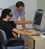 Dr.
Davies and graduate student Mike Swan take a virtual look at a protein
crystal structure.
Dr.
Davies and graduate student Mike Swan take a virtual look at a protein
crystal structure.
So when the Department of Biochemistry and Molecular Biology's ultimate wish to finally establish a structural biology presence with the purchase of a new X-ray crystallography lab and the services of a full-time crystallographer came to fruition, campus scientists and researchers were excited with fervor.
What has been lacking in our research picture was X-ray crystallography, said John Hildebrandt, Ph.D., professor and chairman of the Department of Pharmacology. And it has been somewhat of a deficit within our academic research environment for not having it.
For scientists and investigators, just reaching the stage of identifying proteins involved in a biochemical process and isolating those proteins is a feat in itself often taking many years of exhausting, investigative lab work, according to Hildebrandt.
Nothing gives scientists a bigger jump in defining information than having an X-ray structure, Hildebrandt said, who is conducting research on signal transduction by GTP-binding proteins. It's a quantum leap in understanding about how things fit together. Without these techniques to gather information, interpret data and transcribe it into something visual, your work cannot progress. It's very much like arriving at a wall that you cannot get around.
Academic researchers like Biochemistry and Molecular Biology Associate Professor Eleanor Spicer, Ph.D., has had previous experience working with X-ray crystallography through research collaborations at a distance. Spicer was a member of the 1996 strategic planning committee responsible for bringing crystallography to campus.
As part of her research work on mRNA translation research and gene expression, Spicer would grow protein crystal samples, ship the samples for trials and wait. If nothing happened, the samples and work would ultimately be forgotten. After two years of this, Spicer became extremely frustrated.
Having MUSC crystallographer Chris Davies here means that I can set up my crystal trials growing within my lab and simply walk down the hall and let Chris look at my samples, said Spicer, gratified with the idea that she can now move forward in her work with the presence of X-ray crystallography. Within 20 minutes we'll both know if what I provided is a good protein sample or not. This immediacy will be very helpful.
Once a crystal is identified, Davies uses his skill and expertise to determine the data results. It's a sophisticated, slow process emphasizing the need to obtain good crystal samplesa task not easily met.
For the scientist the work is mostly trial and error but well worth
the struggle because the payoff in science or end-result can be so tremendous,
Spicer said.