Scanner provides non-invasive diagnosis
A new, advanced CT scanner at MUSC is enabling some patients to avoid an invasive cardiac catheterization to diagnose blockage of the coronary artery.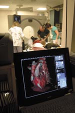 In
the background, from left, CT technologist Kim Strom with David Ball, R.N.,
and CT technologist Robin Brothers prepare a patient prior to scanning.
The monitor in the control room demonstrates the clarity of the images
obtained from the new 64-slice CT scanner.
In
the background, from left, CT technologist Kim Strom with David Ball, R.N.,
and CT technologist Robin Brothers prepare a patient prior to scanning.
The monitor in the control room demonstrates the clarity of the images
obtained from the new 64-slice CT scanner.
The new Somatom Sensation 64-slice computerized tomography system, one of the first five in the nation, gives doctors a view of the coronary arteries not previously possible without the more invasive procedure.
MUSC is one of the first facilities in the nation to have the new scanner, and we have been using it for more than a month, said Philip Costello, M.D., chairman of the MUSC Department of Radiology, who participated in the development of this pioneering technology from its infancy. The only other centers in the U.S. that took delivery of this new technology at the same time as MUSC are the Mayo Clinic, Johns Hopkins, UCLA and the Cleveland Clinic.
 MUSC
radiologist Dr. Bayne Selby provides immediate feedback to patient Doris
Johnson following her scan.
MUSC
radiologist Dr. Bayne Selby provides immediate feedback to patient Doris
Johnson following her scan.
The scanner is located in MUSCs Heart & Vascular Center, and the initial use of the equipment is in the area of cardiovascular medicine. MUSC will be at the forefront of this new technology and will be instrumental in doing comparative studies to determine if the scanner provides just as useful information as catheterization. Costello said that it is appropriate for an academic medical center to work with this highly sophisticated technology and develop protocols for optimal usage.
Imagine the blurry image of a moving race car you would get if you attempted to photograph the moving car with a slow shutter speed, said Christopher D. Nielsen, M.D., director of MUSC adult catheterization laboratories. Compare this with the clarity that could be obtained with an ultra-fast shutter speed.
It is this superior image clarity that helps doctors decide if a patient presenting with chest pains requires medical intervention, treatment with a stent or balloon, or perhaps bypass surgery.
A cardiac catheterization involves the insertion of a small tube into an artery, usually in the groin, and threading it up into the arteries of the heart. A dye is then injected into the coronary arteries, and X-rays reveal the locations and extent of blockages. Nielsen said that the procedure is invasive and involves some risks. The risks are very rare, but they are real, he said. The day of complete elimination of diagnostic cardiac catheterization is not here yet, but I anticipate that this day will come in the next few years as a result of this new advanced technology. For the present, the new diagnostic procedure will be performed on low-risk patients and those for whom catheterization poses a higher than normal risk.
MUSC physicians and researchers plan to use the scanner to follow the results of therapywhether it is stents, laser or surgeryto see if patients are responding to therapy or having recurrences of the initial blockages. Also, lipid-lowering drugs have been shown to decrease plaque in the coronary arteries. This scanner is a means of non-invasively following patients on these drugs to see if the drugs are producing the desired effect.
In addition to providing greater clarity, the new system is more comfortable for the patient. With the older 16-slice CT scanners, patients having their hearts scanned would have to hold their breath for 20 seconds. The 64-slice system scans the heart in only 10 seconds with superior resolution. In addition, for patients with rapid heartbeats, using the older scanner meant having to slow down the heartbeat with drugs. But the most serious limitation of the older scanner is that it only allows visualization of the first two thirds of the coronary artery. As the artery becomes smaller, there is more motion and the image becomes fuzzier. The 64-slice scanner permits visualization of the entire artery.
The speed and high-resolution of the 64-slice system will be especially advantageous for use in children who can be difficult to examine, said radiologist U. Joseph Schoepf, M.D. In six seconds we can scan a childs entire chest and abdomen, looking for congenital deformity of the aorta, he said. The faster scan means a child would less likely have to be sedated.
Another advantage of the 64-slice scanner is in the area of safety,
according to Schoepf. The machine has an automatic adjustment mechanism
that gauges the density of tissue it penetrates and then automatically
adjusts to take the image of using a minimal amount of radiation.
Schoepf brings experience as imaging specialist
by Cindy AbolePublic Relations
Radiologist U. Joseph Schoepf, M.D., knows first-hand the benefits of newer, sophisticated technology especially in the field of interventional radiology and cardiac CT imaging.
 Dr.
U. Joseph Schoepf
Dr.
U. Joseph Schoepf
Schoepf, like his medical colleagues, is impressed with the technical advances in the field of radiology and its many tools used in imaging technology.
As a cardiothoracic imaging specialist, Schoepf works closely with MUSC Heart & Vascular Center cardiologists and other vascular interventional radiologists to refine their imaging skills and develop techniques to create safer, more accurate cardiovascular protocols in the effective diagnosis of coronary artery blockages.
More specifically, Schoepf is among a select field of specialists testing
and evaluating MUSCs new Somatom Sensation 64-slice CT, one of the industrys
most dynamic, non-invasive imaging systems used in the study of heart and
coronary arteries.
MUSC is extremely fortunate to have attracted Dr. Schoepf since his
expertise in cutting-edge research are internationally recognized, said
Radiology Chair Philip Costello, M.D. His efforts will place MUSC Radiology
in the forefront of non invasive cardiovascular research.
In addition to his work with cardiac CT imaging, Schoepf extends his imaging expertise with thoracic surgeons at Hollings Cancer Center. Using newer CT scanning and imaging equipment, Schoepf examines lung chest X-rays and evaluates three-dimensional imaging models to detect early forms of lung cancer in patients.
MUSC offers a wide array of radiological specializations through its many services, said Schoepf, of its interventional radiology, pediatric, neuro and chest imaging specialty programs. Its an advantage when a young, diverse program can offer several imaging specialties to meet every patients needs. Its great for both patient and specialist.
Schoepf came to MUSC last June from Brigham and Womens Hospital in Boston where he worked with Costello. Both collaborated in the clinical and research arenas specializing in pulmonary and thoracic CT research.
As director of CT Research and Development in the Department of Radiology, Schoepf stays busy seeking research topics and developing collaborative partnerships around campus. Currently, he is meeting with MUSCs Digestive Disease Center specialists and the pediatric cardiology staff in the use of modern scanners and equipment to effectively treat patients.
The Austrian native is a medical graduate of Ludwig-Maximilians-University of Munich where he completed his clinical radiology residency. In 2001 he joined Brigham & Womens Hospital as a staff radiologist.
Schoepf is a member of the Radiological Society of North America, Society of Thoracic Radiology, North American Society of Cardiac Imaging and several European professional organizations.
He serves on the editorial board of various scientific journals and is associate editor for cardiac imaging of the journal, Radiology. He has recently published a new radiology text, CT of the Heart: Principles and Application, by Humana Press.
Photos by Margaret Atwood, MUSC Art Services
and Digital Imaging
Friday, Dec. 10, 2004
Catalyst Online is published weekly, updated
as needed and improved from time to time by the MUSC Office of Public Relations
for the faculty, employees and students of the Medical University of South
Carolina. Catalyst Online editor, Kim Draughn, can be reached at 792-4107
or by email, catalyst@musc.edu. Editorial copy can be submitted to Catalyst
Online and to The Catalyst in print by fax, 792-6723, or by email to petersnd@musc.edu
or catalyst@musc.edu. To place an ad in The Catalyst hardcopy, call Community
Press at 849-1778.