3-D Echo views indepth images of heart
by Heather MurphyPublic Relations
MUSCs Pediatric Echo (echocardiogram) laboratory in the Childrens Hospital has acquired a cardiac ultrasound system providing true, real-time, three-dimensional images of the heart.
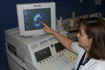 Pam
Ruff demonstrates the depth added to the image of a pediatric patient's
heart.
Pam
Ruff demonstrates the depth added to the image of a pediatric patient's
heart.
Its been our Holy Grail, said Girish Shirali, M.D., pediatric echocardiography director. Because the heart moves so fast, it was difficult to really see depth and see every aspect of a functioning heart with 2-D pictures. Now, weve finally got it.
Cardiovascular disease is the number one killer in the United States, claiming almost as many lives each year as the next seven leading causes of death combined. More than 61.8 million Americans are affected by heart disease, according to current statistics.
Called Live 3-D Echo, this innovation allows cardiologists to view 3-D images of the heart instantaneously, as if they were holding it in their hands, viewing it from different perspectives and looking at the intricate correlation between valves, chambers and vessels.
MUSC has the only four Echos in South Carolina, and is one of only two childrens hospitals in the Southeast with 3-D Echo capabilities.
This real-time view into the heart helps physicians better diagnose cardiac problems. It is becoming a valuable tool in surgical planning, and offers patients a better understanding of their condition.
You can change the angle, look down into the heart, look up through the bottom of the heart, or manipulate the image in just about any way you want, Shirali said. This is wonderful for cardiac surgeons because instead of trying to verbalize everything we see, we can show them amazingly detailed pictures at the same time.
The 3-D imaging makes it possible for surgeons to see the heart from the point of surgical entry, something the 2-D Echos couldnt do.
This machine is wonderful, said Pam Ruff, cardiac sonographer. The probe has 3,000 crystals compared to the old 128 crystal probe and the new 3-D Echos have four Pentium four processors inside. This machine has amazing capabilities.
Unfortunately, the new machine still does not come with anything to help a 3-year-old remain still, Ruff said.
Shirali mentioned the use of sedation could decrease, or once the machines are mastered, time in the Echo lab may be reduced by half for the young patients.
There is currently a revolution in cardiac imaging, one example being the new 3-D Echo, that is allowing the surgeon to have a much better understanding of the anatomy and problems which will be encountered at the time of operative intervention, said Fred Crawford, M.D., Department of Surgery chairman.
Conventional live 3-D imaging (sometimes called 4-D) has been around for almost 10 years for use in other areas like OB/GYN, but was until now it has not been possible to process the high frame rates required to keep up with heart movement. MUSC selected the SONOS 7500 system with Live 3D Echo, manufactured by Philips Medical Systems, to give clinicians the ability to see the heart valves, vessels and chambers in action and slice the image to get progressive views through the heart tissue.
But theres always room for improvement.
We can see a leak, and better than ever before, but we still cant see the actual blood flow itself. Hopefully, well have more progress by June, Shirali said.
These spectacular images from three-dimensional echocardiography
clearly open a new chapter in the care of patients with structural abnormalities
of the heart. The phenomenal flexibility of the imaging direction promises
to provide diagnosticians, interventionalists and surgeons views of the
heart never before obtainable, even by direct visualization when holding
the heart in one's hand.
Phil Saul, M.D.
Director, Pediatric Cardiology
Catalyst Online is published weekly, updated as
needed and improved from time to time by the MUSC Office of Public Relations
for the faculty, employees and students of the Medical University of South
Carolina. Catalyst Online editor, Kim Draughn, can be reached at 792-4107
or by email, catalyst@musc.edu. Editorial copy can be submitted to Catalyst
Online and to The Catalyst in print by fax, 792-6723, or by email to petersnd@musc.edu
or catalyst@musc.edu. To place an ad in The Catalyst hardcopy, call Community
Press at 849-1778.