CAD urged to improve cancer screening
by Dick
Peterson Special to The Catalyst
Nobody’s perfect, not even a computer.
But a study published in CHEST, the Cardiopulmonary and Critical Care Journal, indicates an improved level of detection of possible cancerous lesions of the lung can be achieved if radiologists augment their skills with Computer- Aided Detection (CAD) automated pattern recognition in CT (computed tomography) scans of the chest.
MUSC radiologist U. Joseph Schoepf, M.D., together with Department of Radiology chair, Philip Costello, M.D., led a collaborative study that included researchers from Harvard Medical School and University of Munich, Germany. As a result of the study, five CT scans out of a cohort of 100 interpreted as normal were found to have potentially cancerous lesions, or nodules, on their patients’ lungs.
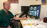 Dr. Joseph Schoepf
displays a CT scan image in which computer assisted detection (CAD)
software found possible cancerous lesions of the lung. The scan was
previously thought to be normal.
Dr. Joseph Schoepf
displays a CT scan image in which computer assisted detection (CAD)
software found possible cancerous lesions of the lung. The scan was
previously thought to be normal.“These were nodules greater than one centimeter in size,” Schoepf said, “and the larger they are the more likely they are cancer.” He said that by the time a nodule reaches one centimeter in size, there is a significantly increased risk that one is dealing with a malignant tumor.
Schoepf said that the study chose 100 CT scans that radiologists had read and found to be free of cancerous lesions. Researchers divided the 100 scans into three groups: patients who had undergone a lung cancer screening, patients who were suspected of having a blood clot in the lung arteries (pulmonary embolism), and patients who had cancer in the past but were thought to be in full remission.
“The research we did used a computer to detect nodules that were missed by human interpretation,” Schoepf said. “We found five patients with lesions greater than one centimeter. The presence of such nodules is certainly something that both the patient and the referring physician would want to know about.”
Schoepf said the study was the first to test CAD in a clinical study involving actual patient care. The study, he said, will enhance interest in CAD use by radiologists in CT scans of the chest.
“We did this with the thought in mind that detection of additional lesions in patients with known lung nodules in most instances does not change patient management, while detection of lesions in scans that were thought to be normal significantly impacts patient management.”
He said that although the study used CT scans already seen by a radiologist, augmenting a radiologist’s inter-pretation with a CAD follow-up would be time consuming and expensive. Running the CAD first followed by a radiologist review risks missing other lesions. Schoepf said that the most efficient approach would use CAD as a system simultaneously with the radiologist’s reading.
As good as the study proved CAD to be, it will never replace the role of the radiologist actually looking at and interpreting the scan. Instead, Schoepf foresees the role of CAD as a valuable tool the radiologist can use to produce more accurate diagnoses.
“Computers see lesions in a way that is different from human perception,” Schoepf said. He explained that regardless of the sensitivity and specificity of a particular CAD tool, CAD is almost always helpful for pointing out lesions that are less conspicuous to human perception.
“We will always need the radiologist to make the judgment call as to whether there is an indication of cancer. As CAD programs develop,” he said, “they will have a greater fit in actual patient care.”
Rapidly becoming a vital part of contemporary medical imaging, CAD’s automated pattern recognition in CT scans of the chest has been preceded by the successful integration of CAD software in other fields, such as the automated evaluation of PAP-smears for early detection of cervical cancer. Schoepf also cited CAD of breast lesions based on X-ray mammography as another example of how the software has enhanced the sensitivity of a medical test in clinical routine.
Previous CAD studies have “resided in the research realm and have been applied to highly selective patient subgroups in an investigational setting.” Schoepf’s current study crossed the threshold from science to practice, he said, and show how CAD tools can enhance diagnosis in thoracic CT.
“We and others are actively working on the development of novel software tools aimed at improving diagnosis of small peripheral pulmonary emboli and coronary artery stenosis in contrast enhanced CT. This way, we hope to outfit physicians with a powerful armamen-tarium for providing their patients with the most accurate and objective diagnosis possible.”
Friday, April 14, 2006
Catalyst Online is published weekly,
updated
as needed and improved from time to time by the MUSC Office of Public
Relations
for the faculty, employees and students of the Medical University of
South
Carolina. Catalyst Online editor, Kim Draughn, can be reached at
792-4107
or by email, catalyst@musc.edu. Editorial copy can be submitted to
Catalyst
Online and to The Catalyst in print by fax, 792-6723, or by email to
catalyst@musc.edu. To place an ad in The Catalyst hardcopy, call Island
Publication at 849-1778, ext. 201.