Doctors explore new ways to diagnose
heart disease
by Mary Helen
YarboroughPublic Relations
Advancing new uses of technology they helped develop, MUSC radiologists and cardiologists are exploring how computed tomographic (CT) scans can now detect vascular blockages.
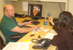
 Dr. Joseph Schoepf
explains to Kathleen Ellis of Business Development and Marketing
Services the new technique he helped develop that helps detect blocked
and impaired blood flow in the heart.
Dr. Joseph Schoepf
explains to Kathleen Ellis of Business Development and Marketing
Services the new technique he helped develop that helps detect blocked
and impaired blood flow in the heart.Using their Dual Source CT scanner, MUSC physicians have developed a new means to analyze how blood flows in the heart in ways that had traditionally relied upon invasive heart catheterization, nuclear medicine and costly magnetic resonance. The technique enables detection of blocked arteries and narrowing of the blood vessels in the heart, as well as poor blood flow in the heart muscle.
The report on the new technique that uses X-ray spectrometry based on CT technology was published in the March issue of the American Heart Association’s Journal “Circulation,” authored by MUSC post doc Balazs Ruzsics, M.D, Ph.D., and co-authored by Heart & Vascular’s U. Joseph Schoepf, M.D., and Eric R. Powers, M.D.
“We are the first in the world to use this technology in a manner that also uses radiation doses that are lower than with the regular Dual Source CT scan,” Schoepf said.
While the CT scan “dissects” the heart into thin layers, enabling doctors to detect diseased vessels and valves, blood flow could not be detected, Schoepf said. So researchers added two X-ray spectrums, each emitting varying degrees of energy, “to gain static and dynamic images of the coronary arteries and the heart muscle,” he said. “On top of that, the use of varying energy levels enables us to see the distribution of the blood in the heart.”
By altering the energy levels of the spectra, radiation doses are kept low, he added.
The device also enables researchers to detect a blockage, to see what the flow is beyond that point, and detect the exact location of the vascular damage.
“We are able to pick up with great clarity any diseased areas of the heart by creating a map of the blood circulating in the heart muscle,” Schoepf said. “We do this by injecting iodine into the patient, and we have determined that where you don’t read iodine in the heart muscle, there is no blood flow.”
For years MUSC has been at the forefront of implementing and refining the latest, most advanced imaging technologies for non-invasively diagnosing heart disease.
If the initial experiences at MUSC are confirmed in larger studies, the technology would enable diagnosing all aspects of heart disease based on a single test that lasts about 15 seconds, with considerable cost-savings. It also would enable greater convenience and reduced radiation exposure for patients, Schoepf said.
In addition to diagnosing the heart, the CT scan also allows doctors to check for other diseases that may be lurking in the lungs or the chest wall, he said.
In their research, the Heart & Vascular Center physicians will systematically compare their new technique to the conventional methods for detecting decreased blood supply in the heart muscle.
Friday, March 14, 2008
Catalyst Online is published weekly,
updated
as needed and improved from time to time by the MUSC Office of Public
Relations
for the faculty, employees and students of the Medical University of
South
Carolina. Catalyst Online editor, Kim Draughn, can be reached at
792-4107
or by email, catalyst@musc.edu. Editorial copy can be submitted to
Catalyst
Online and to The Catalyst in print by fax, 792-6723, or by email to
catalyst@musc.edu. To place an ad in The Catalyst hardcopy, call Island
Publications at 849-1778, ext. 201.